Pet X-Rays
What Are Pet X-Rays?
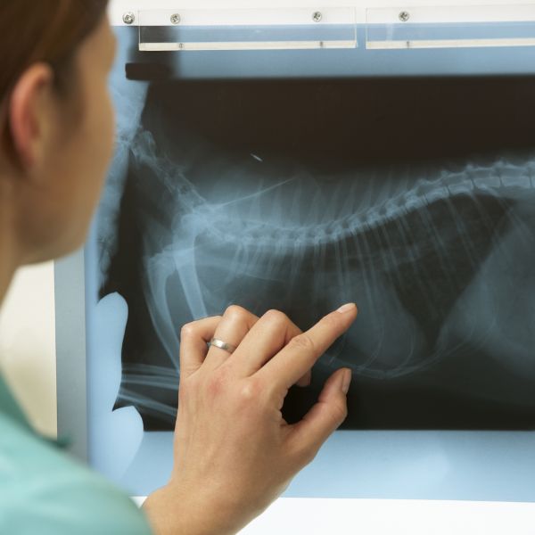
Pet X-rays (radiographs) are a non-invasive imaging technique used to capture images of your pet’s bones, joints, chest, and abdomen.
- Broken bones and fractures
- Joint health and signs of arthritis
- Foreign objects in the digestive tract
- Heart, lung, or abdominal abnormalities
- Tumors, organ enlargement, or fluid buildup
At our hospital, we use digital X-ray technology for sharper images, faster results, and reduced radiation exposure, making diagnostics safer and more efficient.
Why Radiology Matters for Pet Health
Because pets can’t explain their pain or symptoms, X-rays allow us to “see inside” and identify problems we can’t find through a physical exam alone. Radiology supports:
- Fast and accurate diagnoses
- Better treatment decisions
- Surgical planning and post-operative monitoring
- Early detection of disease
Whether your pet has an unexplained limp, persistent cough, or sudden changes in appetite, X-rays help us get answers quickly and compassionately.

Benefits of Digital Veterinary Imaging:
- Immediate image processing and interpretation
- Enhanced clarity for detailed evaluation
- Less stress with shorter exam times
- Safe for pets of all ages and sizes

What to Expect During Your Pet’s X-Ray
For some pets (especially those experiencing pain or who need precise positioning), sedation may be recommended to ensure comfort and safety. Our team uses gentle handling, clear communication, and fear-free techniques throughout the process.
